-
中文名稱:AIFM2兔多克隆抗體
-
貨號:CSB-PA874794LA01HU
-
規格:¥440
-
圖片:
-
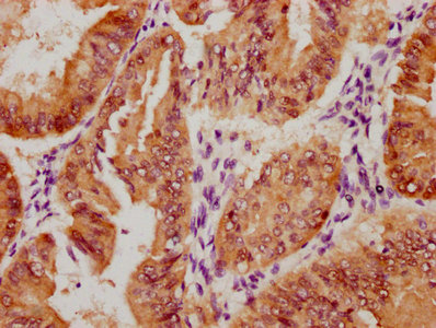
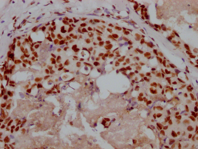
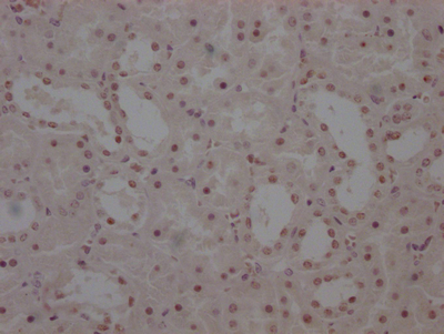
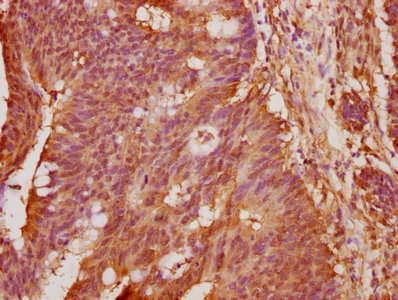 IHC image of CSB-PA874794LA01HU diluted at 1:100 and staining in paraffin-embedded human colon cancer performed on a Leica BondTM system. After dewaxing and hydration, antigen retrieval was mediated by high pressure in a citrate buffer (pH 6.0). Section was blocked with 10% normal goat serum 30min at RT. Then primary antibody (1% BSA) was incubated at 4°C overnight. The primary is detected by a biotinylated secondary antibody and visualized using an HRP conjugated SP system.
 -
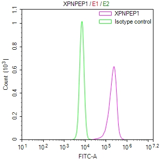
 Western Blot
Positive WB detected in: A549 whole cell lysate
All lanes: AIFM2 antibody at 1:1000
Secondary
Goat polyclonal to rabbit IgG at 1/50000 dilution
Predicted band size: 41, 37 kDa
Observed band size: 41 kDa -
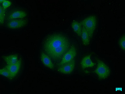 Immunofluorescence staining of HepG2 cells with CSB-PA874794LA01HU at 1:50, counter-stained with DAPI. The cells were fixed in 4% formaldehyde, permeabilized using 0.2% Triton X-100 and blocked in 10% normal Goat Serum. The cells were then incubated with the antibody overnight at 4°C. The secondary antibody was Alexa Fluor 488-congugated AffiniPure Goat Anti-Rabbit IgG(H+L).
-
-
其他:
產品詳情
-
產品名稱:Rabbit anti-Homo sapiens (Human) AIFM2 Polyclonal antibody
-
Uniprot No.:
-
基因名:AIFM2
-
別名:5430437E11Rik antibody; aifm2 antibody; AIFM2_HUMAN antibody; AMID antibody; Apoptosis inducing factor (AIF) homologous mitochondrion associated inducer of death antibody; Apoptosis inducing factor (AIF) like mitochondrion associated inducer of death antibody; Apoptosis inducing factor mitochondrion associated 2 antibody; Apoptosis-inducing factor 2 antibody; Apoptosis-inducing factor homologous mitochondrion-associated inducer of death antibody; Apoptosis-inducing factor-like mitochondrion-associated inducer of death antibody; Cys51Stop antibody; HGNC11998 antibody; p53 responsive gene 3 antibody; p53 tumor suppressor antibody; p53-responsive gene 3 protein antibody; PRG3 antibody; TRP53 antibody; Tumor protein p53 antibody
-
宿主:Rabbit
-
反應種屬:Human
-
免疫原:Recombinant Human Apoptosis-inducing factor 2 protein (110-373AA)
-
免疫原種屬:Homo sapiens (Human)
-
標記方式:Non-conjugated
本頁面中的產品,AIFM2 Antibody (CSB-PA874794LA01HU),的標記方式是Non-conjugated。對于AIFM2 Antibody,我們還提供其他標記。見下表:
-
克隆類型:Polyclonal
-
抗體亞型:IgG
-
純化方式:Antigen Affinity Purified
-
濃度:It differs from different batches. Please contact us to confirm it.
-
保存緩沖液:Preservative: 0.03% Proclin 300
Constituents: 50% Glycerol, 0.01M PBS, pH 7.4 -
產品提供形式:Liquid
-
應用范圍:ELISA, WB, IHC, IF
-
推薦稀釋比:
Application Recommended Dilution WB 1:500-1:2000 IHC 1:20-1:200 IF 1:50-1:200 -
Protocols:
-
儲存條件:Upon receipt, store at -20°C or -80°C. Avoid repeated freeze.
-
貨期:Basically, we can dispatch the products out in 1-3 working days after receiving your orders. Delivery time maybe differs from different purchasing way or location, please kindly consult your local distributors for specific delivery time.
-
用途:For Research Use Only. Not for use in diagnostic or therapeutic procedures.
引用文獻
產品評價

相關產品
靶點詳情
-
功能:A NAD(P)H-dependent oxidoreductase involved in cellular oxidative stress response. At the plasma membrane, catalyzes reduction of coenzyme Q/ubiquinone-10 to ubiquinol-10, a lipophilic radical-trapping antioxidant that prevents lipid oxidative damage and consequently ferroptosis. Cooperates with GPX4 to suppress phospholipid peroxidation and ferroptosis. This anti-ferroptotic function is independent of cellular glutathione levels. May play a role in mitochondrial stress signaling. Upon oxidative stress, associates with the lipid peroxidation end product 4-hydroxy-2-nonenal (HNE) forming a lipid adduct devoid of oxidoreductase activity, which then translocates from mitochondria into the nucleus triggering DNA damage and cell death. Capable of DNA binding in a non-sequence specific way.
-
基因功能參考文獻:
- Low AIFM2 expression is associated with T-cell lymphoblastic lymphoma. PMID: 28534937
- AIF and its family member protein, AMID, are rotenone-sensitive NADH:ubiquinone oxidoreductases (of the NDH-2 type). PMID: 26063804
- HUHS1015 increased nuclear localization of apoptosis-inducing factor-homologous mitochondrion-associated inducer of death (AMID). PMID: 25244912
- induces caspase-independent apoptosis PMID: 11980907
- encodes a homologue of the apoptosis-inducing factor PMID: 12135761
- AMID is a p53-downstream gene involved in tumorigenesis. PMID: 15273740
- Reasults establish a link between coenzyme and DNA binding that likely impacts on the physiological role of AMID in cellular apoptosis. PMID: 15958387
- study of cellular localization of the endonuclease G, AIF & AMID during apoptosis using bioinformatics and image analysis PMID: 17347867
- AIF-M2 lessens survival cell signaling in the presence of foreign (e.g. bacterial and (retro)viral) cytosolic DNA, thus contributing to the onset of apoptosis PMID: 17711848
顯示更多
收起更多
-
亞細胞定位:Lipid droplet. Cell membrane; Lipid-anchor. Cytoplasm. Mitochondrion membrane. Nucleus.
-
蛋白家族:FAD-dependent oxidoreductase family
-
組織特異性:Detected in most normal tissues as two transcripts of 1.8 and 4.0 kb in length, respectively. Highly expressed in heart, moderately in liver and skeletal muscles, and expressed at low levels in placenta, lung, kidney, and pancreas. Both transcripts expres
-
數據庫鏈接:
Most popular with customers
-

-
Phospho-YAP1 (S127) Recombinant Monoclonal Antibody
Applications: ELISA, WB, IHC
Species Reactivity: Human
-
-
-

-
-

-